
Lung injury in broiler chickens is a serious respiratory condition, usually caused by the interaction of multiple factors, such as infections by various pathogens and environmental stress. It has a high mortality rate and causes huge economic losses to the poultry industry. Therefore, understanding the causes of lung injury in broiler chickens and taking effective preventive measures are urgent priorities for the broiler industry.
I. Causes of Lung Injury in Broiler Chickens
The lungs are organs for gas exchange. After entering the lungs, the bronchi do not branch successively into a bronchial tree as they do in mammals. Instead, they form interconnected passages, from whose walls countless tiny pulmonary capillaries (also known as respiratory capillaries) branch off. These are the sites where gas is exchanged directly with the blood in the pulmonary blood vessels. The total respiratory surface area of the pulmonary capillaries per gram of body weight is dozens of times greater than that of mammals, corresponding to the body's intense metabolic needs. Chickens' lungs can also perform double respiration, exchanging gases during both inhalation and exhalation. This greatly increases lung ventilation and improves the efficiency of gas exchange within the lungs. Air sacs are organs unique to birds. They are membranous sacs formed by branches of the bronchi extending beyond the lungs, and most communicate with the internal cavities of many bones.
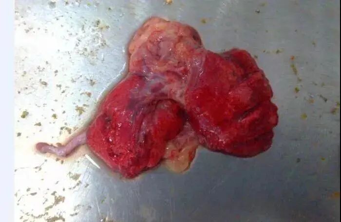
The lungs play an important role in chickens’ physiological activities and are also the organs most susceptible to invasion. Lung damage commonly results from the following causes.
(I), environmental factors1, temperature
1) At low temperatures, cold air irritates the respiratory mucosa and lung tissue, causing local blood vessels to constrict and circulation to be impaired. This leads to inadequate local nutrition, weakening or stopping the activity of the ciliated epithelium, increasing tissue protein breakdown, and consequently increasing large-molecule colloidal proteins in the blood while reducing reticuloendothelial system activity.
This damages the barrier function and consequently reduces the body’s ability to resist infection. The respiratory mucosa becomes susceptible to attack by pathogenic microorganisms, which can spread to the lungs and cause pulmonary infection. Alternatively, the formation of new alveoli may slow, leading to abnormal gas exchange (oxygen and carbon dioxide) and pulmonary hypertension, thereby causing lung damage.
2)At high temperatures, first, chickens breathe faster to dissipate heat, increasing their breathing rate. This causes congestion in the blood vessels of the respiratory mucosa. Over time, vasodilation can easily lead to exudative inflammation; the cilia can easily fall off, exposing and damaging the mucosa, which makes it susceptible to infection by pathogenic microorganisms and promotes the development of pneumonia. Pulmonary congestion progresses to venous congestion and edema. Second, at high temperatures, the heart rate increases. Shallow breathing causes insufficient oxygen in the blood, leading to a compensatory increase in heart rate. The resulting tachycardia can lead to heart failure, which may obstruct venous return and cause pulmonary congestion, edema, and hypoxia in the body.
2, humidity 1)
Excessive humidity: When humidity exceeds 70%, it can affect heat dissipation in larger chickens. To dissipate heat, they breathe faster, promoting heat loss through respiration. High humidity increases pressure inside the lungs, causing congestion in the respiratory organs. Over time, pulmonary congestion can progress to venous congestion and edema; consolidation of the lungs can lead to pneumonia.
2)Low humidity:
Humidity below 30%, dry air, and abundant dust (fine feathers, dirt, powdered materials, etc.) can enter the respiratory system with inhaled air. Over a prolonged period, particles may accumulate in the respiratory tract and adhere to respiratory gland secretions. Excessive accumulation can cause obstruction and difficulty breathing. If large numbers of microorganisms attach to the particles, respiratory or lung infections can also readily occur.
3, negative pressure
The negative pressure in standardized broiler houses should be within −0.08 Mpa. Negative pressure exceeding −0.08 Mpa will cause a degree of lung damage.
When negative pressure exceeds the standard, the airflow speed over the backs of the chickens inside the house exceeds the normal standard, and the amount of oxygen exhausted from the house increases. The oxygen entering the house through the air inlets is insufficient to replenish it to the normal level required by the flock. Oxygen deprivation causes pulmonary vasoconstriction and increases pulmonary capillary venous pressure. On the other hand, oxygen deprivation stimulates the release of large amounts of catecholamines, causing systemic vasoconstriction and increased total peripheral resistance, increasing left ventricular afterload, altering left ventricular function, and reducing ejection fraction. At the same time, the higher absolute value of the elevated intrapulmonary negative pressure leads to greater negative pressure across the myocardium, while prolonged oxygen deprivation causes myocardial hypoxia throughout the heart; both can also alter left ventricular function. These factors can all trigger pulmonary edema, followed by a series of lung injuries.
4, harmful gas levels exceed the standard
In the chicken house, the concentration of harmful gas ammonia is greater than 20 mg/cubic meter, and the concentration of hydrogen sulfide is greater than 15 mg/cubic meter. These gases can irritate mucous membranes. Damage to the respiratory mucosa causes the cilia to fall off, facilitating pathogens’ invasion of the lungs. When the carbon dioxide concentration is greater than 2500 mg/cubic meter, gas exchange is affected, causing pulmonary hypertension and thereby lung damage.
(II) Pathogen infection
Common pathogens that cause lung infections include: Escherichia coli, staphylococci, mycoplasma, molds, H9, infectious bronchitis, etc.
1, Escherichia coli pneumonia
The basic pathological feature of Escherichia coli pneumonia is microcirculatory disturbance in the lungs. Due to increased capillary permeability, large amounts of fibrinogen exude into the alveoli, causing extensive consolidation of the lung tissue. In the early stage of the lesion, the lung lobes are congested and edematous, and large amounts of serous exudate are present in the alveolar cavities. Pulmonary carnification: When the exudate cannot be completely absorbed and cleared due to a low number of phagocytes or impaired phagocyte function, it is organized by granulation tissue, and the lung tissue at the lesion site becomes brown, fleshy fibrous tissue.
2, staphylococcal pneumonia
The lungs of chickens infected with staphylococci are dark purple, with pale yellow, millet-sized fibrinous nodules on the surface and within the lung tissue.
3, Mycoplasma damage to the lungs
Mycoplasma gallisepticum often causes complications together with other microorganisms. Escherichia coli is one of the microorganisms that commonly occurs in conjunction with Mycoplasma. Newcastle disease and infectious bronchitis, among others, can promote Mycoplasma infection. When all three occur together, they cause a severe air sac infection and also cause lung infection.
4, damage to the lungs caused by infectious bronchitis
Infectious bronchitis virus infection in chickens 18h can cause respiratory cilia defects, rounding and sloughing of mucosal epithelial cells, and infiltration by small numbers of heterophils and lymphocytes. Catarrhal exudate is present in the trachea or bronchi of affected chickens. In severe cases, caseous plugs may be seen in the posterior trachea or bronchi, and pneumonia of varying extent may be observed around the large bronchi.
5, lung damage caused by chicken mold
The lungs and trachea turn blackish-purple or grayish-white and become hardened. The cut surface shows necrosis, the air sacs are cloudy, and there are fungal nodules.
6, low-pathogenicity influenza (H9) causes damage to the lungs
Low-pathogenicity avian influenza in chickens (such as H9N2) affects the respiratory system. Its characteristic lesions include catarrhal, fibrinous, serofibrinous, mucopurulent, or fibrinopurulent inflammation. The tracheal mucosa is congested and edematous, with occasional hemorrhage; it also causes inflammation of the lungs to varying degrees.
II. Preventive measures
1, improve the environment
During production, manage conditions in strict accordance with the actual environment and standards for temperature, humidity, and ventilation, providing the flock with a comfortable, healthy growing environment. In flocks kept in an environment with an ammonia concentration of 10 mg/L or 20 mg/L, tracheal mucus secretion increases, the cilia surfaces become rough, and mucociliary movement deteriorates. The ability of the air sacs, liver, and lungs to eliminate Escherichia coli decreases. If colibacillosis pneumonia occurs, the flock becomes more susceptible to infectious bronchitis and Newcastle disease. This demonstrates the importance of the environment for the flock.
2, develop a reasonable immunization program
Develop a reasonable immunization program based on the actual conditions in the breeding area, and make every effort to ensure that antibody levels against common diseases remain above the normal standard during the corresponding antibody troughs, so that the flock can safely pass through these troughs and grow healthily.