
Epidemiological characteristics of avian pneumovirus
Chickens and turkeys are natural hosts of this disease; pheasants, guinea fowl, and quail can also be infected. The virus is transmitted mainly through contact, and sick and recovered birds are the main sources of infection. Contaminated water sources and feed, personnel, equipment, and the movement of infected or recovered birds can also spread it. Airborne transmission has not been confirmed, while vertical transmission may occur.
Clinical symptoms
Clinical symptoms vary greatly depending on factors such as feeding and management practices and the presence or absence of complications.
Clinical symptoms in young chickens after infection: tracheal rales, sneezing, nasal discharge, frothy conjunctivitis, swelling of the infraorbital sinuses and subcutaneous edema of the neck; in severe cases, coughing and head shaking.
Clinical symptoms in laying hens after infection: breeder chickens and laying hens usually become ill at peak egg production, with egg production declining by 5%-30%, and sometimes by 70%; severe cases can result in oviduct prolapse. Eggs have thin, rough shells, and the hatchability of hatching eggs decreases. The disease course is generally 10-12 days. Some individuals also show respiratory symptoms such as coughing. Egg quality is also affected, and the disease often occurs as a mixed infection with infectious bronchitis and Escherichia coli. In addition to head swelling, specific neurological symptoms are also observed. Apart from some sick chickens showing only extreme depression and coma, most cases exhibit disorientation, with manifestations including head shaking, torticollis, ataxia, unsteady gait, and opisthotonos. Some chickens tilt their heads upward in a “star-gazing” posture. Sick chickens are reluctant to move, and some die because they do not eat.
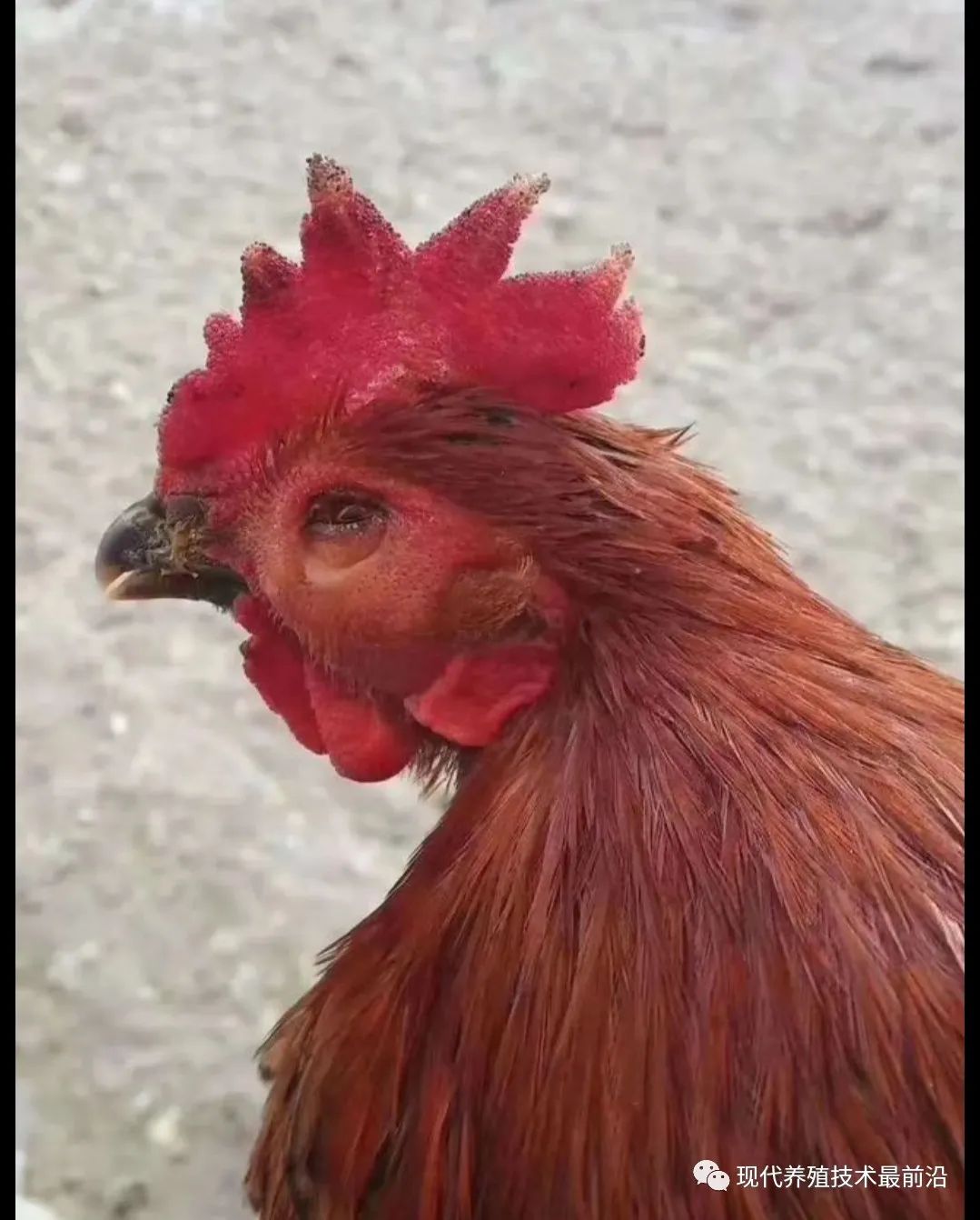
Clinical symptoms of swollen head syndrome caused by pneumovirus: broiler chickens are often affected at 4~5 weeks of age. The infection rate can reach 100%, and the mortality rate varies, ranging from 1%~20%. The first symptom is sneezing. Within one day, conjunctival redness and swelling of the lacrimal glands occur. Over the following 12~24 hours, subcutaneous edema begins to appear on the head, first around the eyes, then spreading to the rest of the head and subsequently to the submandibular tissues and wattles. In the early stage, chickens scratch their faces with their claws, indicating localized itching. They then become depressed, reluctant to move, and have reduced appetites. They may show enlarged infraorbital sinuses, torticollis, ataxia, opisthotonos, and generalized respiratory symptoms.
Clinical signs of viral airsacculitis caused by avian pneumovirus: difficulty breathing, neck extension and open-mouth breathing, coughing, secondary colibacillosis in later stages, increased mortality, and even loss of the entire flock.
Pathological changes
The lesions found at necropsy are not specific to this disease, and mixed infections with avian Bordetella, Ornithobacterium rhinotracheale, and Mycoplasma are common, which can lead to misdiagnosis. Congestion of the nasal turbinate mucosa appears first, followed by widespread red to purple discoloration. Histological changes include flattening of the epithelium, gradual loss of cilia, congestion and lymphoid hyperplasia beneath the epithelium and in the lacrimal glands, and an increase in plasma cells in the lacrimal glands. Head swelling in affected birds is caused by subcutaneous edema and expansion around the eye sockets. Severe cases show cyanosis and swelling of the wattles. The subcutaneous tissue of the head develops yellow edema that may progress to suppuration, and the eyelids close due to edema and conjunctivitis. In broilers, lesions mainly present as airsacculitis, pericarditis, pneumonia, and perihepatitis. In laying hens infected after the onset of lay, watery or mucoid exudate is present in the nasal cavity two weeks after infection, tracheal mucus increases, and there may be yolk peritonitis, deformed yolks, and even degeneration of the ovaries and oviducts, concentrated egg white, solidified yolk, and folded shell membranes in the oviduct. As the disease progresses, tracheitis and pulmonary necrosis may develop. Histological examination reveals accumulations of lymphocytes and heterophils in edematous areas.
Diagnosis
Virus isolation
SPF chicken embryo or turkey embryo isolation method: Inoculate the yolk sac or allantoic cavity of 6-7-day-old chicken or turkey embryos. After 3-5 passages, the virus may cause impaired embryonic development, bleeding on the embryo’s surface, and even death.
Tracheal ring culture method: Culture tracheal ring tissue from 18-day-old chicken embryos; ciliary growth stops.
Cell culture: chicken and turkey kidney cells, chicken embryo liver or fibroblast cells, and VERO cells. After viral propagation, the virus can be identified using electron microscopy, neutralization tests, ELISA, indirect fluorescent antibody techniques, and other methods.
Bacterial isolation can detect the presence of Escherichia coli, Haemophilus, Mycoplasma, Bordetella, and others.
Serum antibody measurement: measured using ELISA, the indirect fluorescent antibody test, virus neutralization tests, and other methods.
Immunofluorescent staining of diseased tissue.
Animal inoculation test: inoculate susceptible chickens and turkeys with diseased tissue; the corresponding swollen-head symptoms and lesions should be reproduced.
Control measures
The quality of husbandry and management has a major impact on the infection and spread of this disease. Factors such as poor temperature control, high stocking density, poor-quality litter, inadequate hygiene, mixing birds of different ages, and failure to recover fully after disease infection can all trigger pneumovirus infection. Beak trimming or vaccination during a non-safe period may increase the severity of pneumovirus infection and increase mortality.
Strengthen husbandry and management: Carefully and consistently implement the husbandry and management system. In addition, good biosecurity measures are key to preventing pneumovirus from entering the farm.
Hygiene management measures: Strengthen disinfection protocols, rotate disinfectants with different active ingredients, improve poultry-house hygiene, reduce stocking density, lower the concentration of ammonia in the air, and maintain good ventilation in poultry houses. These measures can help prevent or reduce the occurrence of disease and its severity.
Prevent secondary bacterial infections: Antibiotics may be used for treatment, while also increasing vitamins and electrolytes.
Immunization: Where vaccines are available, vaccination may be considered. Develop a reasonable immunization program based on the vaccine instructions and the actual circumstances of your flock. Live vaccines may be considered for commercial chicks and broilers, and inactivated vaccines for layer chickens.