
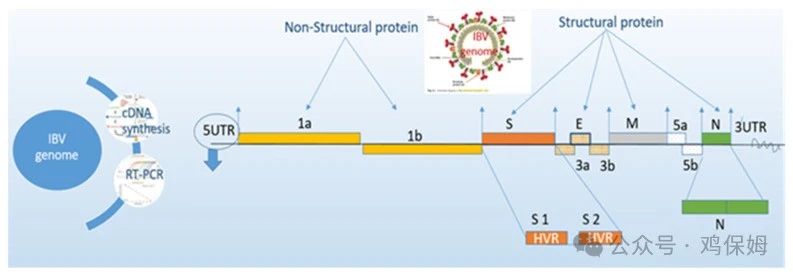
Few methods are available for diagnosing infectious bronchitis (IB) to identify the virus in current infections or the specific antibody response following viral infection. As described below, selecting the best test for successfully detecting avian infectious bronchitis virus (IBV) is difficult and confusing because of various factors.
1. Clinical signs
The clinical signs of IBV resemble those of other respiratory infections, but some distinctive lesions are part of strain variation and are difficult to distinguish from those of other respiratory viral infections. Although clinical signs are matched according to the profile, serological studies have never been matched against anatomical lesions.
Serological titers occasionally show an increasing trend, but this does not correspond with the clinical signs; therefore, confirmation of IBV is problematic. Consequently, confirmatory diagnosis is based on accumulated data in farm records, including egg quality and production curves, mortality, seroconversion or antibody levels, vaccination reports, and blood serology, with clinical signs in IBV-infected chickens matched simultaneously against PM lesions. Samples for IBV isolation are best collected immediately, while clinical signs are evident. RT-PCR for reverse-transcribed RNA is an effective method for detecting IBV in active infections. Some studies indicate that nutritional deficiencies are an important consideration in detecting IBV.
2. Vaccination
Vaccination is a major factor affecting IBV identification because the infecting IBV vaccine strain must be distinguished from other virulent strains. In unvaccinated flocks, a field challenge can be detected simply by RT-PCR, RT-qPCR, or positive IBV titers several weeks later. Therefore, identifying a field challenge or active infection in vaccinated flocks is very difficult because antibodies circulate in the blood. Today, vaccine strains can be distinguished from field strains using DIVA (differentiating infected from vaccinated animals) technology with inactivated oil-emulsion vaccines. They can be detected by simple neutralization tests, enzyme-linked immunosorbent assay (ELISA), and indirect immunofluorescence assays.
3. Test selection
The choice of diagnostic technique is very important for accurate diagnosis in relation to sample collection and the number of samples. Advanced ELISA tests can distinguish between antibodies produced by vaccination and those produced by infection.
Antibody titers tend to rise abnormally high 2-4 weeks after infection; over the following weeks, titers gradually decline slowly. The next blood sample should then be collected 2 weeks later, but no later than 4 weeks, from the same infected house, because titers are delayed in reaching their highest level as the immune system needs time to develop immunity.
Viral concentrations in infected tissues are highest within 3-5 days after infection; molecular assays and virus isolation should be selected instead of serological monitoring. At present, common molecular methods have been used for IBV diagnosis, particularly RT-PCR, but IBV isolation is more challenging and time-consuming, and can only be carried out from infected chickens because multiple passages in embryonated eggs are required before the virus replicates. Alternatively, because mutations frequently occur at primer-binding sites, various types of variants may evade detection, so primers need to be updated frequently for use in molecular assays.
4. Serology
The mean titer provides information about the antibody response of the birds tested within a flock after vaccination. The mean titer should fall within a predictable range, with a uniform distribution of mean titers in a normal flock and inadvertently higher titers in a diseased flock.
One study showed that the antibody pattern induced by vaccination should not be disrupted by field infection. After infection, the geometric mean titer (GMT) should increase abnormally when the titer level is at least 2 times higher than the level expected after vaccination, or when the mean titer level is at least 2 times higher than before infection. Sometimes, mean titers gradually rise after vaccination; however, no clinical signs are observed during normal production, indicating good vaccine protection and a better response.
According to general parameters, the coefficient of variation (CV) percentage after inactivated vaccination remains below 40%, and below 60% after live vaccination, indicating good vaccination uniformity. When CV is less than 20%, titers show good uniformity, but this may indicate previous disease exposure or good vaccination. Therefore, cross-checking against mortality, clinical signs, and production records is necessary to ultimately confirm IBV. A good assessment of the CV percentage is significantly below the expected level after vaccination or significantly below the level in early infection cases. Therefore, a CV% below 30% after a single dose of live IBV vaccine is designated as a suspected challenge. In contrast, after vaccination with a highly immunogenic IBV variant vaccine, the CV percentage is usually below 45%. In this case, before administering the inactivated vaccine, a series of multiple live vaccinations is first used to promote 100% seroconversion in birds, which is the main principle affecting titer persistence rather than CV percentage measurements.
5. Organ selection and sample quality
The upper respiratory tract (UTI) is the primary site of IBV replication; subsequently, viremia leads to viral multiplication and spread through the bloodstream to other tissues. Due to continuous cross-infection in infected or immune flocks, IBV can persist for a long time in the cecal tonsils and kidneys. In acute cases, clinical signs are found in the respiratory tract, making it the preferred site for pooled samples. In addition, kidney, cecal tonsil, and cloacal samples are selected in chronic infections in chickens vaccinated during the laying phase, while only small amounts of virus can be predicted in the respiratory tract. Collected organs should be refrigerated as quickly as possible to preserve viral viability for further molecular diagnosis. If freezing or refrigeration facilities are unavailable, samples should be preserved in 50% glycerol for several days.
6. Immunity and challenge during infection
Serological monitoring can directly help assess the immune status of poultry, viral challenge, and the duration of viral infection. Different types of commercial ELISA kits use different cutoff values and mathematical formulas to calculate and convert ELISA results. The most common practice in agriculture for enhancing immunity to control IBV infection is to first administer a live attenuated vaccine, followed by an inactivated vaccine with a normal titer level to provide good protection against IBV.
IBV challenge typically presents with respiratory signs when unusually high IBV titers are checked 6-8 weeks after infection. For breeder chickens, the normal guideline for calculating challenge in infected chickens is that the average post-infection titer is at least 2 or 3 times the expected vaccination titer (baseline), as shown in Figures A and B. For broilers, challenge typically presents with unusually high IBV titers, and the average post-infection titer should exceed 3-4 K (Idexx standard) after live vaccination with normal serology, as shown in Figures C and D, respectively.
In ELISA testing, diagnosis should be based on expected vaccination titers, taking into account the vaccination program, the geographic distribution of IBV, outbreak history, the specific vaccine, and the type of poultry being examined. The combination of these respective factors with regular flock reports is used to determine whether serological results are normal or abnormal. Many factors affect antibody levels, particularly the virus strain, as well as factors related to the chicken (e.g., breed, type, age, immune status, and nutrition), the vaccine (e.g., attenuation level, dose, live or killed vaccine, and storage conditions), and the vaccination schedule (e.g., correct schedule, preparation, and route of administration).
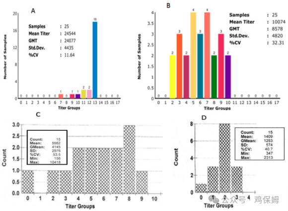
Figure 2 compares IBV serological analyses at 26 weeks in (A) a challenged flock and (B) a normal flock in two broiler-breeder houses. The average titer of the challenged flock was calculated to be three times that of the normal flock, and the coefficient of variation (CV%) was too low compared with previous serological tests using a Symbiotic test kit in the United States. The high challenge titers following IBV infection at peak lay were almost all in a single bar. In broilers, (C) the high challenge titer following IBV infection observed at 35 days of age exceeded 3-4 K, calculated after IBV infection, compared with (D) normal serology, based on the standard for the Idexx ELISA test kit in the United States.
7. Genetics and immunosuppression
Genetic traits have a substantial effect on resistance and susceptibility to IBV. Some experimental studies have shown that different genetic traits, mortality rates, viral growth, and variable histopathology were found in different breeding lines after IBV immunization. However, because susceptibility differs among chicken lines, there are some differences among testing methods. Some immunosuppressive infections are associated with worsening IBV disease. For example, infectious bursal disease (IBD), Marek’s disease (MD), inclusion body hepatitis (IBH), and chicken infectious anemia (CIA) may suppress immune status at a young age. This can disrupt acquired vaccine immunity to IBV by stimulating lymphocyte depletion in the bursa and thymus, leading to greater resistance to challenge. The main damage is affected by subclinical infections, making immunosuppressive agents difficult to understand.
Laboratory diagnostic methods used to confirm IBV infection involve other respiratory viruses, which require continuous serological monitoring to determine the actual source of the current infection. Similarly, cyclosporine is classified as a T-cell immunosuppressive drug, can significantly reduce immunity in poultry, and is highly stimulated in IBV infections.
Source: Chicken Nanny, Author: Md. Safiul Alam