
Editor's note: While organizing past documents, we came across this article. Although opinions vary widely on proventriculitis and ventriculitis in chickens, comprehensive, valuable articles are rare. This article systematically describes the history and development of proventriculitis and ventriculitis, and is undoubtedly a beacon for industry professionals and technical staff working to solve problems. We would like to thank the original presenter, Mr. Stephane Lemiere (Merial Animal Health Co., Ltd.), China Poultry, and Mr. Li Wanmeng and Ms. Li Bei of Merial for their hard work.
1, functions of the gizzard and proventriculus:
The proventriculus is olive-shaped and varies greatly in size. Its chief cells secrete gastric acid, lowering the pH of the stomach contents and enabling pepsin to break down proteins. Mucosal cells secrete mucus to protect the epithelial cells on the stomach surface from erosion by gastric acid and to prevent food from rubbing against them.
The gizzard has two muscle layers, a thick layer and a thin layer. The koilin layer serves as a protective layer, and its color is related to the color of bile. If bile refluxes, the koilin layer will appear green. The gizzard's main function is mechanical digestion; it has no secretory glands and therefore produces no enzymes. The koilin layer can withstand mechanical pressure and protect the gizzard cells. The gizzard mainly grinds food. During digestion, it withstands considerable pressure, and the koilin layer ensures that this pressure does not damage the gizzard. When the gizzard is examined during a postmortem, small stones or coarse sand may be found inside; these can enhance the gizzard's digestive function. This shows that good feed processing helps improve flock performance; poor feed processing can cause indigestion in the gizzard, thereby affecting the chickens' production performance.
The proventriculus, gizzard, and duodenum are very important for digestion in chickens.
2, the digestion process of feed:
The proventriculus is an organ containing secretory glands, equivalent to the stomach in other animals, and produces gastric juice containing gastric acid and digestive enzymes. Food moves from the proventriculus to the gizzard, an organ with a hard koilin layer and muscle layers. In the gizzard, food is ground and thoroughly mixed with gastric juice.
3, proventriculitis and gizzard erosion:
In fact, when proventriculitis or gizzard erosion is found during poultry necropsies, field veterinarians will suspect that the feed has been contaminated with fungi and mycotoxins. Of course, when these problems are encountered clinically, they may not necessarily receive sufficient attention from the poultry farm. Laboratory testing is rarely performed to confirm the cause, and many companies will not take the issue seriously unless production performance has already been affected. In fact, proventriculitis and gizzard erosion are not solely issues involving fungi and mycotoxins; other combined factors may also be involved.
Proventriculitis is not very common in clinical cases, and proventriculitis caused by viral infection is also relatively uncommon. However, proventriculitis is associated with infections by many viral diseases (such as Marek’s disease virus, reovirus, infectious bursal disease virus, Newcastle disease virus, avian influenza virus, avian reticuloendotheliosis virus, and infectious bronchitis virus, among others).
Overall, the main causes of proventriculitis are bacterial infections and viral diseases. Proventriculitis is closely associated with gizzard erosion, which is generally considered to be caused by the following factors: mycotoxins and biogenic amines; and infectious diseases, including infections with reovirus, fowl adenovirus, oncogenic viruses, Cryptosporidium, Newcastle disease virus, avian influenza virus, and infectious bursal disease virus, among others.
When discussing proventriculitis, we are not referring only to a problem in an individual chicken. A group of chickens, a poultry house, or a poultry farm should be considered as the subject of study when assessing the occurrence of proventriculitis.
3.1 Proventriculitis: Proventriculitis is characterized by inflammation and swelling of the proventriculus and thinning of its wall, and is common mainly in broilers. Proventriculitis is a relatively troublesome disease for integrated poultry slaughtering companies. This is because proventriculitis obstructs the passage of food through the digestive tract, increasing the risk of proventricular rupture during slaughter and, consequently, the rejection rate during the slaughter process. In production, inflammation of the proventriculus impairs its function and affects feed digestion, which can lead to a reduction in feed conversion efficiency10%.
Necropsy of a proventriculus affected by proventriculitis may reveal swelling and inflammation of the inner mucosa accompanied by edema, thereby affecting the secretory function of the proventriculus. This reduces the secretion of digestive enzymes and affects feed digestion.
The main causes of proventriculitis include mycotoxins and biogenic amines in feed, and infectious diseases such as viral arthritis, reovirus, fowl adenovirus, infections with oncogenic viruses such as Marek’s disease virus, Newcastle disease virus, and avian influenza virus. Immunosuppression caused by infectious bursal disease virus is also a very important factor contributing to proventriculitis in chickens. However, the relationship between immunosuppressive diseases and proventriculitis is relatively subtle and easily overlooked. Of course, proventriculitis is also closely associated with gizzard erosion.
When the proventriculus is infected with pathogens, histological examination may reveal hypertrophy of the proventricular epithelial cells, as well as lymphocyte proliferation and infiltration, indicating deep non-suppurative necrotizing proventriculitis, accompanied by symptoms such as hemorrhage and edema on the inner surface of the proventriculus. Sometimes, lymphoid tumors can be seen in cases of proventriculitis during necropsy. Virulent Newcastle disease virus can cause hemorrhagic proventriculitis. In the early stages of avian influenza, necropsy of the proventriculus may reveal hemorrhage and inflammation on its inner surface.
Proventriculitis is not very common in production. However, when broiler proventriculi are examined at necropsy, proventriculitis is sometimes found (see Figure 1). If cases are only sporadic, they are generally not caused by infectious viral factors. However, when large-scale cases of proventriculitis are found in a flock, viral causes of the proventriculitis often need to be investigated.
Infectious viral proventriculitis (see Figure 2) is an important factor in reduced broiler production performance. It can greatly reduce body-weight uniformity in broilers and cause poor feed digestion, affecting growth. To date, the causative agent of infectious viral proventriculitis has not been identified. However, previous studies considered the causative agent to be a novel, previously unrecognized double-stranded RNA virus (RII/3 virus).
RII/3 virus was isolated from the proventriculus of chickens infected with infectious viral proventriculitis. The virus, after laboratory passage, can reproduce the disease.
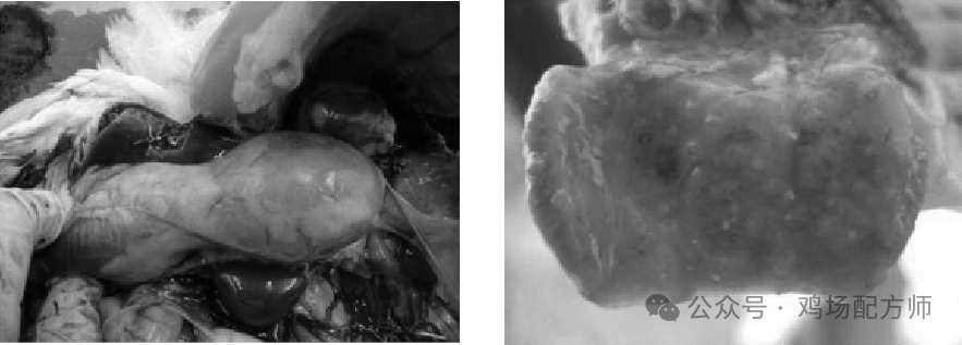
Figure 1: Proventriculitis in 28-day-old broilers. Figure 2: Viral proventriculitis.
Infectious viral proventriculitis can damage 80% of the pepsinogen- and hydrochloric acid-secreting cells, leading to poor feed digestion, a low feed conversion rate, slow growth, and other effects in chickens.
3.2 Gizzard erosion:
3.2.1 Causes of gizzard erosion: The specific pathogens responsible for this disease have not yet been identified. The most common reports link it to toxins in feed. However, lesions have also been found in 1-day-old chicks that had not eaten feed. Known factors in gizzard erosion include biogenic amines in feed. Biogenic amines can cause decarboxylation, disrupt the catabolism of amino acids in feed, and cause poisoning in chickens. The most common substances found in feed of animal origin are histamine, 3-hydroxytryptamine, 5-hydroxytryptamine, histidine, gizzard eroding factor, and serotonin in the blood. These factors may lead to gizzard erosion.
3.2.1.1Histamine: Histidine in fish meal can be converted into histamine by the action of several types of bacteria. Histamine can bind to cell receptors and stimulate the production of hydrochloric acid. This lowers the pH value, causing erosion of the surface of the gizzard.
3.2.1.2Gizzard erosion factor: Overheating fish meal and improper storage can both produce gizzard erosion factor, which is 10 times more toxic than histamine.
3.2.1.3Biogenic amines: In addition to causing gizzard erosion, biogenic amines can also cause proventriculitis and hyperplasia of the proventricular glands, leading to reduced flock performance.
3.2.1.4Mycotoxins: Mycotoxins such as T2, 二醛酸蔍草镰刀菌烯酮, and serpentines can cause gizzard erosion (see Figure 3) as well as oral erosion and intestinal bleeding. Citrinin can cause gizzard rupture, while cyclopiazonic acid can cause proventricular swelling, dilation of the proventricular neck, and mucosal necrosis. Mycotoxins are difficult to manage. In production, compliant feed should be used, and flock management should be strengthened. Mycotoxin-induced damage to the gizzard mucosa is shown in Figure 4.
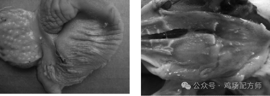
Figure 3: Gizzard erosion caused by mycotoxins Figure 4: Gizzard mucosal damage caused by mycotoxins
3.2.1.5 Viral infectious gizzard erosion: In laboratory assessment of a field strain, serotype one fowl adenovirus (FAV-99ZH) can cause gizzard erosion in poultry (see Figure 5). Gizzard erosion and pancreatitis lesions can be found 3 to 4 days after infection.
Gizzard erosion is also associated with infection by serotype 8 fowl adenovirus (FAV - MOI3 and G0054).
Histological examination may reveal degeneration of the koilin layer, mucosal necrosis, intranuclear inclusion bodies, and inflammatory cell infiltration.
3.2.1.6Stress: Stress has been shown to cause gizzard erosion and several other clinical symptoms in chickens. Clinical trials show that stress caused by cold or hot weather reducing flock water and feed intake can lead to gizzard and proventricular lesions (erosion, ulcers, and hemorrhage). Good poultry-house management can address stress.
3.2.1.7Other potential factors: Other potential factors include amino acid imbalance, excessive sulfates, lack of fiber in the feed, insufficient feed and water, excess vitamin A, vitamin E deficiency (see Figure 6), and incubation stress.
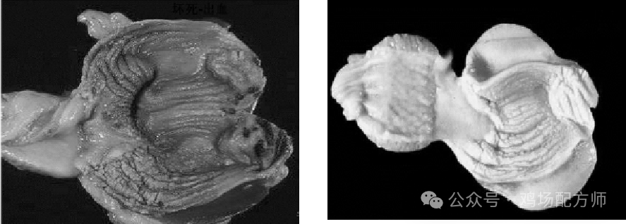
Figure 5: Gizzard erosion caused by viral infection Figure 6: Gizzard erosion caused by vitamin deficiency
3.2.2 1 days of age, gizzard erosion: Necropsy of gizzard erosion at 1 days of age shows localized to extensive hemorrhage and necrosis in the inner layer of the gizzard mucosa (koilin layer). Lesions observed at the hatchery appeared before the chicks started eating. The rough, red mucosa indicates that the hemorrhage progressed from the inside outward. Histological examination revealed pinpoint to extensive hemorrhage and sloughing of epithelial cells in the koilin layer. Microscopic findings indicate that the lesions occurred 2–3 d before hatching; gastric hemorrhage during incubation is associated with stress.
4, conclusions and recommendations:
Glandular gastritis and gizzard erosion are problems on poultry farms with high-density, intensive farming and deserve our attention. Management factors and infectious disease factors can cause these problems, leading to reduced flock production performance and economic losses. It is not sufficient to assume that mycotoxins may be the sole cause of glandular gastritis and gizzard erosion; a definitive diagnosis must be made.
Teamwork among production staff and managers, clinical veterinarians, and nutrition experts is essential. Effective monitoring of feed mill processing and close monitoring of mycotoxins and animal-derived additives can reduce the occurrence of these problems. Standardized hatchery management can reduce cases of gizzard erosion in 1-day-old chicks. Biosecurity measures are key to controlling infectious factors.
Note: Reprinted from: Chinese Journal of Poultry Science, Volume 2010, Issue 32 of 15. The original article was compiled by the editor based on conference recordings and slides, with some content omitted, and was not reviewed by the author. This is hereby noted.
Source: Poultry farm formula specialist; compiled by Zhao Junhe