
Looking back at the prevalence of chicken diseases in China over the past few years, the disease with the highest popularity index is “chicken Mycoplasma synoviae disease.” The approximate history of its spread in China is as follows: it occurred sporadically in broiler chickens around 2011 and soon became widespread; it began occurring sporadically in layer chickens in 2013, but spread relatively slowly, and did not become widespread until 2014. To date, its prevalence and rate of spread continue to show an upward trend, making it one of the few diseases with a relatively long period of prevalence. Most chicken diseases generally follow a pattern: “broilers first, layers later”—that is, the disease spreads first among broilers, and only 1-2 years later among layers. In addition, in China, a particular disease (or strain) generally remains prevalent for approximately 2-3 years. Mycoplasma synoviae is still showing an increasing trend.
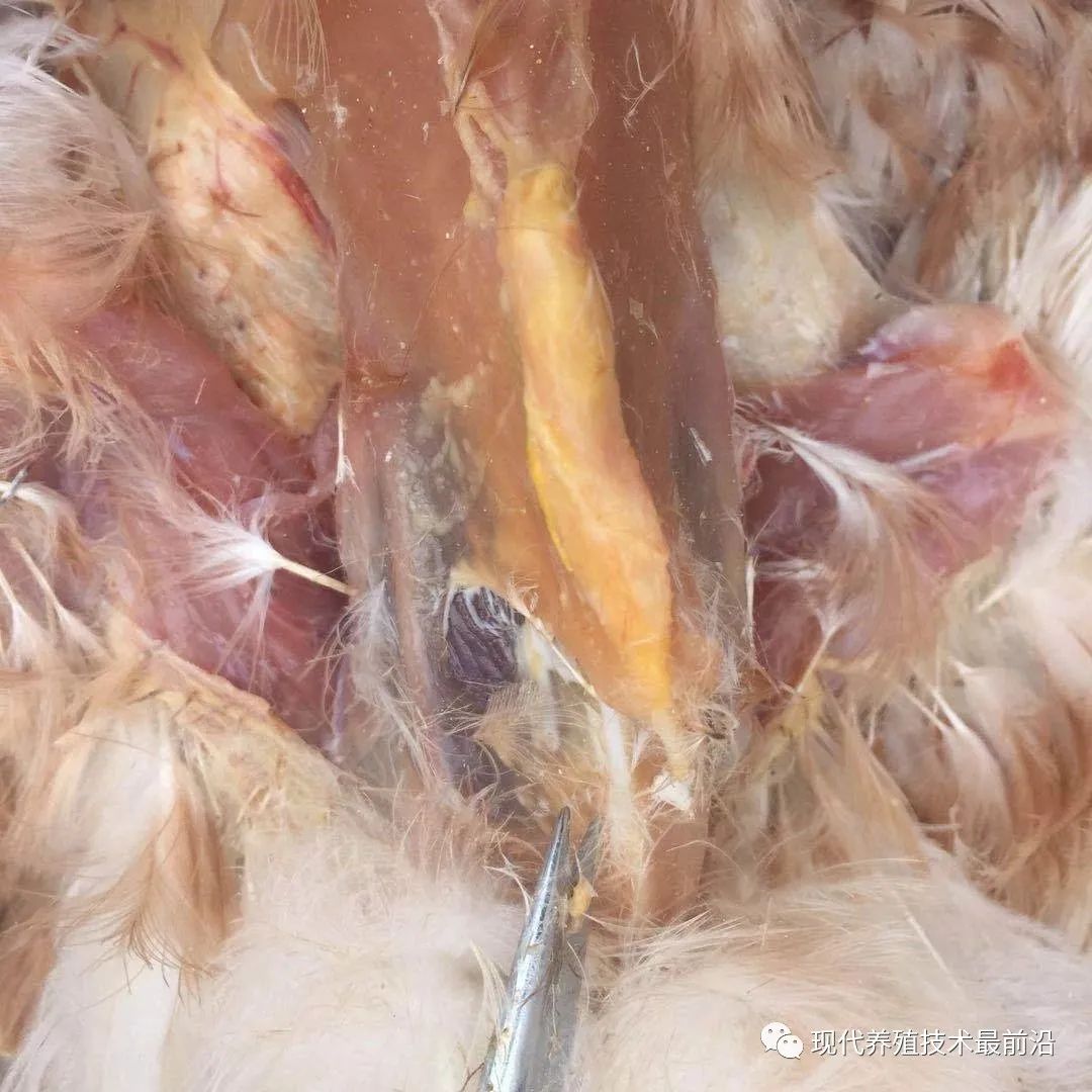
Because this disease progresses slowly and runs a long course, once a flock becomes infected, eradication is very difficult. As a result, it can persist in flocks for a long time, leading to low feed utilization, slow growth and development, higher culling rates, and reduced egg production. When the pathogen is present in a flock, mixed infections can occur readily, aggravating the disease and increasing mortality, resulting in serious economic losses.
What exactly is this disease, described as having “such tenacious vitality and such vigorous reproductive capacity”? What causes it, and how can it be prevented and controlled?
I. What is chicken synovitis?
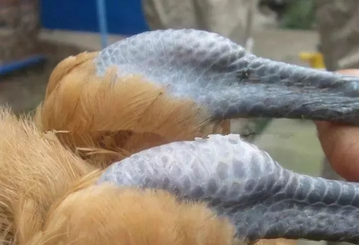
Chicken Mycoplasma synoviae disease is an infectious disease of young chickens caused by Mycoplasma synoviae. It is also known as infectious synovitis of chickens. Chickens older than 12 weeks rarely develop the disease; it most commonly affects chickens aged 9-12 weeks. The morbidity rate is 5%-10%, and the mortality rate is generally below 10%, though in severe cases it can reach about 75%. Affected chickens may have shrunken, pale combs; separate themselves from the flock; prefer to lie down; tuck their heads in and close their eyes; grow slowly; have rough, disheveled feathers; walk with a slight splayed-leg gait; limp; and become anemic. They may pass green feces, develop ascites, and become emaciated. The areas around the joints are often swollen, especially the hocks and toes; the swelling can sometimes reach the size of a pigeon’s egg and feel fluctuant to the touch. In the late stage of the disease, the joints become deformed, and the birds may be unable to rise after lying down for a prolonged period. Although they have an appetite, they become extremely emaciated because they cannot eat, and eventually die from exhaustion or complications from other diseases. Egg production in hens may decrease by 20%-30%.
Mycoplasma synoviae is a microorganism intermediate between bacteria and viruses, with two main characteristics:
1, very fragile outside the chicken:
①Under normal conditions, it can survive in the air for only two days.
②It is very sensitive to any disinfectant or most antibiotics; even soapy water can kill it.
③Its in vitro culture requirements are very demanding, so producing a truly effective vaccine is costly.
2, very resilient inside the chicken:
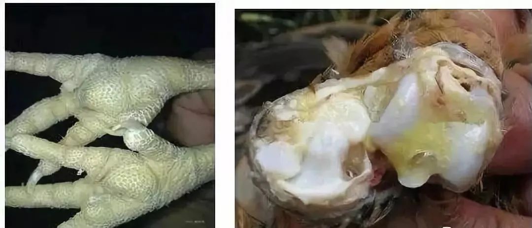
The main affected sites are the keel, hock joints, and footpads. These areas have poor blood circulation, making it difficult for many drugs to reach them through the bloodstream. Therefore, once infected, Mycoplasma synoviae is very persistent.
二、What is the disease progression?
The progression of Mycoplasma synoviae disease in chickens has three steps: First, the pathogen is present; second, it enters the chicken; third, it reaches the target organ.
First, if there is a pathogen, where does it come from?
The three main sources of Mycoplasma synoviae:
(1) Breeder flock contamination: vertical transmission to chicks.
(2) Vaccine contamination: vaccines contaminated with Mycoplasma synoviae.
(3) Environmental contamination: the pathogen is present in the affected farm area itself, or has been transmitted from another source.
Second, how does it enter the chicken’s body?
As the saying goes, “Disease enters through the mouth.” The three main entry routes for Mycoplasma synoviae are:
(1) Respiratory route: ① Pathogens in the environment enter the respiratory system directly through airborne transmission; ② contaminated live vaccines enter the respiratory system directly or indirectly during intranasal, ocular, drinking-water, or aerosol administration.
(2) Digestive tract entry: Contaminated material is pecked and enters the digestive system, such as feces from sick chickens or feed pecked by sick chickens.
(3) Bloodstream entry: ① Vertical transmission: Parent breeder chickens that are diseased or carry the pathogen transmit it through the bloodstream, allowing it to enter the chicks directly; ② Contaminated syringes, needles, or wing-web inoculation tools enter the bloodstream directly during immunization.
Third, reaching the target organs—which organs?
The main target organs affected by Mycoplasma synoviae are the keel, hock joints, and footpads.
III. What causes the disease?
The causes of Mycoplasma synoviae disease are:
First, there must be both a real culprit and an accomplice; neither can be absent. The real culprit is the Mycoplasma synoviae pathogen, and the accomplice is any factor that reduces the flock’s resistance.
Second, when the combined attacking force of the real culprit and the accomplice exceeds the flock’s resistance, the flock becomes ill.
Third, the rapid development of the pullet industry may have contributed to the spread of MS. Transportation and the stress of adapting to a new environment reduce the flock’s disease resistance: it is undeniable that, like Mycoplasma and Escherichia coli, MS is an opportunistic disease. The large-scale, long-distance, cross-regional transport and sale of pullets not only expand the scope of disease transmission, but long-distance transport during the 4–16 weeks, when acute disease is most likely to occur, is bound to promote the disease’s occurrence (some flocks show no disease at the pullet farm but become ill after arriving at the farmer’s premises). Inability to adapt to a new environment and new cages also affects flock health.
Fourth, high-density, intensive production favors the spread of MS. Compared with traditional individual brooding, pullet farms generally keep larger flocks at higher stocking densities. Under these conditions, even a slight lapse in management or inadequate biosecurity can allow a “spark” to become a “raging fire.”
Four. How great is the impact of the disease?
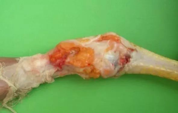
Direct impact: Affected birds experience pain or paralysis due to keel cysts, swollen joints and footpads, and inflammation of the synovial bursae and tendons. This in turn affects feed intake, resulting in poor body weight and uniformity. The disease is chronic and debilitating. Because affected birds fall significantly short of target body weight, the culling rate is 5-20%; the disease rarely causes death directly. Mortality increases significantly only when colibacillosis occurs as a secondary infection. In such cases, the direct cause of death is Escherichia coli, not Mycoplasma synoviae.
Indirect impact: It affects body weight and uniformity, thereby indirectly affecting the egg production rate, by approximately 5-10%.
Five. How is the disease diagnosed?
Synovitis diagnosis has two purposes: to accurately diagnose affected birds and determine whether they carry the pathogen.
1, Is it easy to diagnose affected birds?
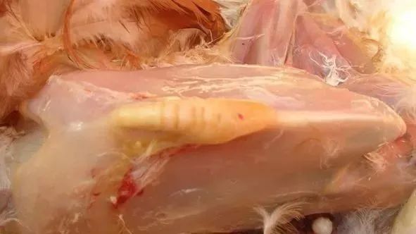
At necropsy, most cases in the early stages of the disease show marked swelling of the joints and tendon sheaths, with a viscous, cheese-colored to grayish-white exudate present. In cases with a longer disease course, the exudate is caseous. The surfaces of infected joints are often yellow or orange-red. Characteristic exudate is more abundant in the hock joints, wing joints, or footpads. The joint membranes are thickened, and the joints are enlarged and protruding.
Histopathological examination: Heterophils and fibrin infiltration can be seen in the joint cavities and tendon sheaths, especially those of the toe joints and hock joints. The synovial bursa membranes show villous formation, with nodular infiltration and proliferation of lymphocytes and macrophages in the sub-synovial layer.
Hematological examination: The total red blood cell count and hemoglobin level are decreased, and abnormal-shaped and abnormal-staining red blood cells appear. The total white blood cell count is markedly increased; lymphocytes and eosinophils are decreased, while heterophils and monocytes are increased.
2, How can you determine whether a bird is carrying the pathogen?
① Infected chickens, whether or not they recover, carry the pathogen for life. This is now a consensus, but the pattern of pathogen shedding is still under study.
② Chickens that have not shown disease need to be tested for the pathogen regularly to determine scientifically whether they are carriers. Throat or cloacal swabs are generally collected. Testing methods include PCR or pathogen isolation and culture. PCR testing is faster and relatively less costly, while pathogen isolation and culture are more difficult to perform and more costly.
Six. How can it be effectively prevented and controlled?
It must be emphasized that mycoplasma synoviae disease has persisted for 8 years. It has not only remained uncontrolled, but has also shown a trend toward accelerating spread, whereas other poultry diseases can generally be effectively controlled in 2-3 years. This clearly shows that it is difficult to control this disease using just one or two simple methods. An effective prevention and control plan must be developed scientifically and rationally based on the characteristics of mycoplasma synoviae, the disease progression and causes, and the specific characteristics of the farm.
1, General approach
The general approach is: “eliminate the true culprit + control the accomplices”
2, What methods are there to eliminate the true culprit?
(1) Eliminating the true culprit outside the body: Make full use of the fact that Mycoplasma synoviae is “very fragile outside the chicken.” Isolation, cleaning, disinfection, drying, and other methods are all effective ways to eliminate the true culprit outside the body.
(2) Eliminating the true culprit inside the body: vaccination + medication
Age in days for the first preventive medication = earliest age of susceptibility - number of days needed to achieve an effective blood drug concentration
Number of preventive medication courses = period of susceptibility ÷ number of days the effective blood drug concentration is maintained
3, What should be noted when controlling the accomplices inside the body?
Why did prevention fail despite using many preventive medicines? The reason is ineffective control of the contributing factors. The four common contributing factors are poor ventilation, heat and cold stress, immune stress, and poor-quality feed.
To control the contributing factors, two points need to be addressed:
First, reduce the likelihood of contributing factors occurring and lessen their impact.
Second, use functional medicines as an adjunct at special times to strengthen the chickens’ constitution.
Strengthen husbandry management
Provide a suitable rearing environment and maintain good ventilation while preventing the flock from getting chilled. Reduce stress, adopt an all-in/all-out rearing system, increase the interval between batches, and strengthen disinfection and monitoring to reduce the likelihood of this disease occurring.
Strengthen litter managementThe severity of Mycoplasma synoviae disease is usually proportional to inadequate litter management. After the disease occurs, poor litter management can greatly increase the culling and mortality rate. Therefore, litter management should be strengthened to prevent damp litter from causing high ammonia concentrations in the poultry house, which can trigger Mycoplasma synoviae infection.
Control mold and mycotoxins
Controlling mold and mycotoxins and improving the body's immunity are effective measures for preventing Mycoplasma synoviae.