
Clinically, it is most difficult to distinguish mild avian influenza from Newcastle disease, especially after mixed infection, when control is difficult. The treatments for these two diseases are completely different. Once a misdiagnosis delays the best time for treatment, it becomes very difficult to control deaths, particularly in broiler chickens.
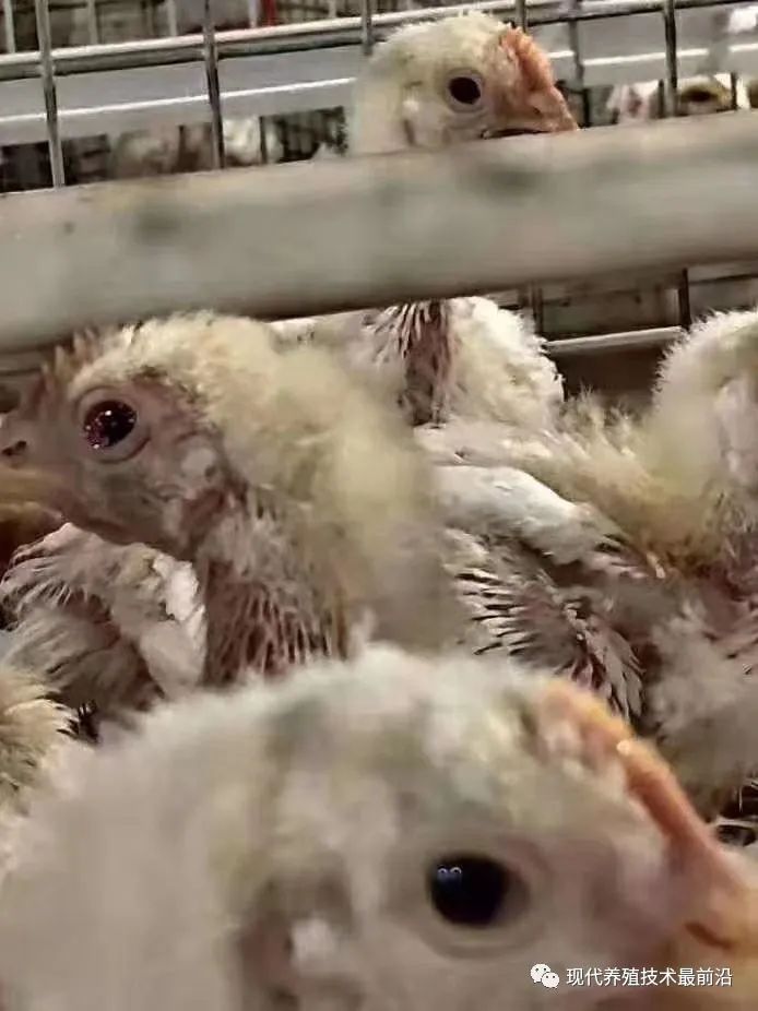
Different clinical presentations
1, clinically, a few grower chickens with Newcastle disease suddenly die at first, followed by respiratory symptoms, coughing, difficulty breathing, and elevated body temperature. After 2—3 days, some chickens develop neurological symptoms: head and neck tremors, trembling, nodding, leg paralysis, and weight loss, with an increase in deaths.
Newcastle disease does not cause head swelling; avian influenza does. Avian influenza typically presents with elevated body temperature, depression, reduced appetite, coughing, sneezing, rales, difficulty breathing, watery eyes, ruffled feathers, a hunched posture, swelling of the head and face, and cyanosis of the skin (comb and wattles). Newcastle disease is mainly characterized by a wet cough, while avian influenza is mainly characterized by rattling breathing and watery eyes. Avian influenza causes a runny nose, whereas Newcastle disease does not.
Newcastle disease mainly kills thin chickens, which die lying prone; avian influenza mainly kills fat chickens, which die lying on their backs (the trachea is obstructed by yellow, dry, fibrous material, and yellow, dry, fibrous material adheres to the lung surface. Some thin chickens may also die; there may be pericarditis and perihepatitis, very thick air sacs, and mottled kidneys). Head and facial swelling may occur, along with screaming, coughing, and rattling breathing. Avian influenza may cause hemorrhage in the scales on the legs; Newcastle disease does not.
In mild avian influenza, the initial symptom is diarrhea; in the middle and later stages, the feces are yellow, green, or a mixture of yellow and green. In the later stage, some chickens pass loose, orange-yellow feces; this is an important characteristic. Newcastle disease causes severe inflammation of the crop; when the chicken is held upside down, mucus flows from the throat and has a sour, foul smell, whereas this does not occur with avian influenza. Avian influenza develops rapidly and may easily recur soon after recovery. Newcastle disease generally does not recur after recovery and the development of immunity.
2, distinguishing the diseases by autopsy findings
Whether in Newcastle disease or mild influenza, hemorrhagic spots appear in the thymus. The changes caused by Newcastle disease are only in the front 1-4 pairs of thymus glands, H7. Mild influenza generally causes hemorrhage or enlargement to appear first in the lower pairs of thymus glands; in the early stage, hemorrhagic spots and enlargement generally appear first in the lower thymus glands. There is no need to examine the thymus in laying hens, because it has already largely regressed by sexual maturity (the thymus is largest at around 60-80 days of age and gradually shrinks afterward).
Changes in the cecal tonsils. In chickens with Newcastle disease, the cecal tonsils invariably show severe hemorrhage and enlargement. If it is H7 mild influenza, some cases cause severe hemorrhage in the cecal tonsils, some cause less severe hemorrhage, and a large proportion do not cause cecal tonsil hemorrhage.
Changes in intestinal lymphocytes
1. Chicken lymph nodes are generally located in the lower part of the duodenum and have a strip-like, irregular island-like shape (2-3 centimeters long). In the absence of Newcastle disease or influenza, they are white and latent, with no distinct islands visible. After either Newcastle disease or mild influenza occurs, the islands become distinct, with more than 1 obvious hemorrhagic spots on them; mild influenza is more often characterized by diffuse hemorrhage, which is less common in Newcastle disease.
2. Within 2-6 centimeters behind the yolk stalk, lymphoid arcs are present and are normally latent. Changes here generally occur only with Newcastle disease; influenza usually does not cause them, or does so not at all. The main finding is an arc-shaped structure about half the size of a soybean. In typical or atypical Newcastle disease, it only protrudes, with 1 or more hemorrhagic spots on it, or with hemorrhage over the entire area. With virulent Newcastle disease, severe ulceration occurs, covered with a sticky green substance. In cases of mild influenza with severe mortality, a white outline occasionally becomes visible. Please note this.
3. Changes in the ileal lymphoid arcs. The changes seen here in mild influenza are the same as those behind the yolk stalk; Newcastle disease also causes changes consistent with those behind the yolk stalk.
4. The narrow section of the cecum. Mild influenza causes no changes here or no diffuse hemorrhagic spots, whereas Newcastle disease is characterized by 1 or more millet-like raised spots with hemorrhage, ranging in size from small millet grains to rice grains. Their size is related to the chicken’s age, the severity of the disease, and the severity of the hemorrhage. Please consider this carefully. To emphasize, changes in the rectal mucosa are not a diagnostic basis for ND or AI and can be ignored clinically. The rectal mucosa generally has densely packed pinpoint hemorrhagic spots (commonly seen in renal bronchitis) and linear hemorrhage (seen in Newcastle disease).
5. H7 mild influenza can also cause fibrinous peritonitis, which appears yellow. In the early stage, laying hens also show only fibrinous peritonitis; only later does yolk peritonitis appear. However, Newcastle disease generally does not cause peritonitis, although mild yolk peritonitis may occur in the later stage.
6, changes in the trachea, bronchi, lungs, and air sacs. Newcastle disease typically causes pin-point hemorrhages on the laryngeal cartilage and hemorrhage in the tracheal rings, without severe congestion. In contrast, avian influenza causes severe hemorrhage throughout the trachea, with abundant mucus and yellow, dry, fibrinous material blocking the trachea. Yellow, dry, fibrinous material may also adhere to the lung surface. Some emaciated chickens may die; the heart and liver may be surrounded by fibrin; the air sacs may be very thickened; and the kidneys may be swollen and mottled.
7, changes in the proventricular papillae. Newcastle disease generally causes hemorrhage at the tips of the papillae, while mild avian influenza more commonly causes hemorrhage at their bases. Hemorrhage may also occur in some intermediate papillae. In some cases, the entire proventricular mucosa becomes reddened and hemorrhagic.
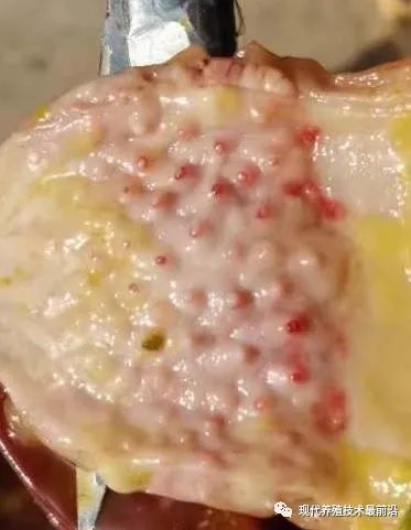
8, influenza can cause hemorrhage in the subcutaneous fat of the neck, abdominal fat, and coronary fat, whereas Newcastle disease generally does not. Influenza can cause splenomegaly with white necrotic spots, whereas Newcastle disease generally does not.
9, Newcastle disease can cause mucus and pin-point hemorrhages in the bursa of Fabricius. Influenza can cause swelling of the bursa, with mucus and hemorrhage. Influenza can also cause hemorrhage in the leg and breast muscles, whereas Newcastle disease does not. At this point, it must be distinguished from infectious bursal disease; otherwise, injecting infectious bursal disease antibodies may cause extensive mortality.
Clinically, influenza is mainly characterized by swelling of the head and face, which is not seen in Newcastle disease. Take care to distinguish it from the following conditions:
1, distinguishing it from infectious coryza. H9 Mild avian influenza causes swelling of the head and face that appears firm and pale. The wattles are generally not swollen on their own, or are not swollen. In contrast, infectious coryza causes pale-red edema that feels fluctuant or soft when pressed, and is accompanied by swollen wattles. Some people say that influenza also causes swollen wattles. I would say that this is the wattle becoming raised due to firm swelling at its base caused by influenza. In addition, infectious coryza invariably causes crusting around the nostrils and nasal discharge, among other signs, so it should not be difficult to distinguish.
2, distinguishing it from swollen-head colibacillosis. The swelling caused by swollen-head colibacillosis is exactly the same as that caused by mild avian influenza. However, swollen-head colibacillosis does not cause respiratory abnormalities; even when they occur, they account for 0.1%. It also does not cause nasal discharge. However, unaffected chickens in the flock continue to eat and drink well, and egg production and egg color remain good. Among affected chickens, unless the corresponding anti-colibacillosis medication is injected, each one found will die. Only the affected chickens pass white, watery droppings that cover a relatively large area. A mixture of ampicillin and gentamicin injected into these chickens produces a clear effect after 2 doses (penicillin generally should not be mixed and injected with gentamicin, as this reduces efficacy). Administering a high-grade anti-colibacillosis medication to the flock quickly brings the outbreak under control.
Gross lesions and clinical distinctions
In terms of anatomical changes, in addition to the changes described above in H9 mild influenza with H7, there are also changes associated with severe septicemic Escherichia coli, with severe fibrinous covering of the heart, liver, and peritoneum. In fact, this is not caused by colibacillosis. So why should treatment with Escherichia coli medication be given as well? I believe it is because severe inflammatory changes have already developed in the internal organs, so antimicrobial treatment is necessary as an adjunct.
So, how can it be distinguished from septicemic form of fowl cholera? There is only one distinguishing feature: the “three inflammations” caused by influenza do not smell foul, whereas those caused by E. coli have a foul smell. If this change is observed, first carefully check whether there are one or more chickens in the flock with swollen heads or faces; then check for obvious respiratory symptoms. If both are present, it is mild influenza.
In addition, with mild H9 influenza, there are cord-like fibrinous lesions under the skin of the neck; influenza can cause redness and swelling in the kidneys, with slight urate deposits in the ureters. Newcastle disease does not cause these changes. In the intestines, influenza causes 1 or more hemorrhagic spots about the size of millet grains, which closely resemble those caused by coccidiosis. However, when lesions caused by this disease are scraped with scissors, there is an obvious feeling of bleeding, which is absent in Newcastle disease.
mixed infection
Newcastle disease is generally accompanied by mixed infections with infectious bronchitis, chronic respiratory disease, infectious bursal disease, coccidiosis, and Escherichia coli. Avian influenza is generally accompanied only by co-infection with Escherichia coli, which worsens the condition and causes high mortality.