
In recent years, the incidence of cutaneous and visceral hemangioma disease in laying hens has risen markedly. In some areas, it is often accompanied by “big liver disease,” seriously affecting egg production rates and increasing mortality and culling rates. Mortality in some affected flocks can reach 20%–30%, causing substantial economic losses to the poultry industry.
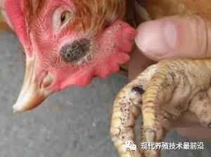
I. Clinical signs
Affected chickens are lethargic, with shrunken, yellow combs and a one-finger gap between the legs. Examination of the skin and feathers reveals blood clots ranging from the size of mung beans to that of Chinese dates on the skin of the chest, neck, toes, and tail. The surrounding feathers are stained with blood.
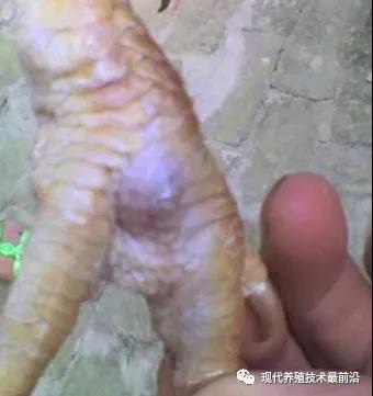
II. Necropsy findings
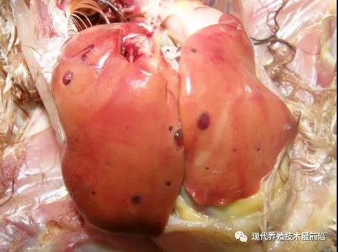
Some chickens that died of the disease have scattered or densely clustered dark red blood blisters in the skin and subcutaneous tissue. In some, scattered or densely clustered dark red blood blisters are found on the surface and within the substance of internal organs, including the conjunctiva, liver, lungs, spleen, stomach, and kidneys. Chickens with visceral hemangioma may sometimes have blood clots in the abdominal cavity. In some cases, hemangiomas of varying sizes are found on the surfaces of the membranes of the sternum, neck muscles, and leg muscles; in the deep layers of the leg muscles; and in the thoracic and abdominal air sacs, ovaries, intestines, mesentery, oviducts, oviduct mesentery, uterine wall, and uterine mucosa. Some chickens with hemangioma disease also show typical lesions of “big liver disease”: the liver, spleen, and kidneys are extremely enlarged, and numerous grayish-white tumor lesions of varying sizes are present on their surfaces and cut surfaces.
III. Causes
Hemangioma disease in laying hens, like the more commonly seen avian lymphoid leukosis (that is, “big liver disease”) and osteopetrosis, is caused by avian leukosis/sarcoma group viruses (ALV). The virus is transmitted vertically when it passes from the hen’s ovary or oviduct into the egg, or horizontally to chicks hatched from virus-free breeding eggs. The chicks then shed the virus in their feces throughout their lives, while the virus persists in their organs and tissues. Some chicks carrying the virus develop and grow normally, but some develop tumorous lesions before the onset of laying.
Four. Control measures
1. Control the introduction of breeding stock. It is not realistic for grandparent and parent stock farms to eliminate this disease. Breeding stock introductions must be strictly inspected; do not introduce chicks or eggs from primary breeding farms where this disease is prevalent or that have not been cleared of it. In addition, hatching eggs must be collected, stored, and incubated under strict disinfection procedures, especially fumigation.
2. Maintain good hygiene and disinfection to prevent early infection. Because susceptibility to this disease is high and it can spread horizontally, particular care is needed: especially after early infection, birds may carry and shed the pathogen for a long time or permanently, and eventually die. Chicks and adult chickens must be raised separately to prevent cross-infection. Improve husbandry and management, reduce stress as much as possible, and strengthen the birds’ resistance. In addition, prevent and control other diseases, strengthen disinfection of syringes, and during vaccination ensure that the needle is changed after each chicken, to strictly prevent transmission of this disease through injection or blood collection.
3. Develop a disease-free, cleared breeding flock. At present, there is neither an effective vaccine to prevent this disease nor an effective drug treatment. Since transmission is mainly vertical, the most fundamental preventive measure is to develop a breeding flock free of this disease.