
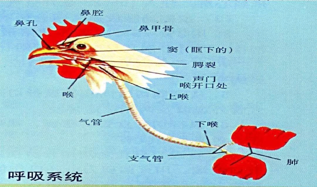
The chicken respiratory system includes the nasal cavity, larynx, trachea, bronchi, lungs, and air sacs.
(1) Nasal cavity
The chicken’s nasal cavity is short and narrow, and is divided by the nasal septum into left and right cavities. Each nasal cavity contains 3 cartilaginous nasal conchae, and connects posteriorly to the pharynx through the choanae. The nasal cavity is not only a passage for incoming air, but also cleans, warms, and moistens the air.
(2) Larynx and trachea
① The larynx is supported by the arytenoid and cricoid cartilages.
The glottis is a longitudinal slit. The mucosa around it has papillae that help prevent food from accidentally entering the trachea. Intrinsic laryngeal muscles are distributed between the laryngeal cartilages and function to open and close the laryngeal opening.
② The trachea is a long, tubular structure made up of numerous complete cartilage rings. It can expand and contract, allowing flexible movement of the neck. The trachea runs along the ventral side of the neck, slightly to the right. After entering the thoracic cavity, it divides into two bronchi that enter the lungs. At the point of bifurcation is a specialized posterior larynx (syrinx), the chicken’s vocal organ.
The trachea is also an important site for regulating body temperature through evaporative cooling. Therefore, in hot weather, chickens breathe with their mouths open and at an increased rate.
(3)The lungs are the organs of gas exchange.
After entering the lungs, the bronchi do not branch successively into a bronchial tree as they do in mammals. Instead, they form interconnected passages, from whose walls countless tiny pulmonary capillaries (also called respiratory capillaries) branch off. These are the sites where gas exchange occurs directly with the blood in the pulmonary blood vessels.
Calculated per gram of body weight, the total respiratory surface area of the pulmonary capillaries is dozens of times greater than that of mammals, matching the body's high metabolic demands. Chicken lungs can also perform double respiration: gas exchange takes place during both inhalation and exhalation, greatly increasing lung ventilation and improving the efficiency of gas exchange within the lungs. Air sacs are organs unique to birds. They are membranous sacs formed by bronchial branches extending beyond the lungs, and most connect with the internal cavities of many bones.
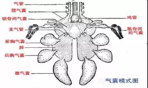
Chickens have 9 air sacs, with a large volume accounting for approximately 85% ~90% of the total volume of the respiratory organs, which is 5 ~7 times the volume of the lungs. Therefore, the air sacs can store more air and act like bellows during respiration, driving gas to pass through the lungs twice, during inhalation and exhalation. This means that gas exchange can take place in the lungs during both inhalation and exhalation, adapting to the body’s intense metabolism.
How can chicken diseases be diagnosed based on lesions in the lungs and air sacs?
(1) Yellow, millet-sized to pea-sized nodules in the lungs: seen in chick aspergillosis; also seen in tuberculosis in adult chickens.
(2) Grayish-black or pale-green mold patches on the lung surface: seen in aspergillosis in young or adult chickens.
(3)Pulmonary congestion and edema: seen in fowl cholera, avian streptococcosis, septicemic pullorum disease in chicks, infectious bursal disease in chickens, and Escherichia coli septicemia in chickens; also seen in leucocytozoonosis in chickens and cottonseed meal poisoning.
(4)Pulmonary granulomas: seen in the pneumonic form of pullorum disease in chicks and Escherichia coli disease in chicks; also seen in chickens infected with air sac mites.
(5)Pulmonary tumor nodules: seen in the visceral form of Marek’s disease in chickens.
(6)Blood clots in the lungs: seen in leucocytozoonosis in chickens.