
In growing chicks, a dietary deficiency of calcium or phosphorus can cause abnormal skeletal development, even when the diet supplies adequate vitamin D3. A deficiency of calcium or phosphorus prevents normal bone calcification. Rickets is seen mainly in young poultry, while calcium deficiency in laying hens leads to a decline in eggshell quality, followed by osteoporosis. This condition, caused by the depletion of bone structure, has commonly been called “caged layer fatigue syndrome” since it was first identified around 60 years ago, when cage systems were introduced. The condition is rare in non-cage systems, but this phenomenon interacts with reduced egg production. Over time, excessive mobilization of calcium from the bones to compensate for insufficient dietary calcium erodes bone structure, eventually leaving it unable to support the hen’s body weight.
1 Rickets
Rickets is most common in young meat poultry, and its main characteristic is inadequate bone mineralization. Although diets deficient in or imbalanced in calcium, phosphorus, or vitamin D3 can also cause the problem, cellular calcium deficiency is the primary cause. Young broilers and turkey poults show signs of lameness at about 10-14 days of age. Their bones are soft and pliable, their ribs are flattened, and bead-like enlargements form at the vertebral junctions. Affected birds have disorganized cartilage matrix, irregular vascular permeability, and signs of metabolic disorders involving collagen precursors such as hyaluronic acid and desmosine.
Rickets is not caused by an abnormal onset of bone mineralization, but by disruption of the early maturation process. Symptoms include frequent enlargement at the ends of long bones and widening of the growth plates. Determining whether rickets is caused by calcium, phosphorus, or vitamin D3 deficiency, or by excess calcium (leading to phosphorus deficiency), may require analysis of blood phosphorus levels and examination of parathyroid function.
In most cases of rickets, vitamin D3 deficiency is generally suspected. This may be due to a simple deficiency of vitamin D3 in the diet, inadequate potency of the D3 additive, or other factors that reduce the absorption of vitamin D3.
The best measure for preventing rickets is to provide adequate levels and potency of vitamin D3 additives and optimize the utilization of fat-soluble compounds in the diet formulation. Young poultry have a limited ability to digest saturated fatty acids. The undigested portion readily binds with calcium to form insoluble soaps, resulting in secondary calcium deficiency. This condition cannot be diagnosed by testing dietary calcium; it must be confirmed through fecal mineral analysis.
The diet must maintain a precise balance between calcium and available phosphorus. Therefore, particular caution should be exercised when using ingredients with highly variable calcium and phosphorus contents, such as animal protein.
In recent years, 25 (OH)₂D₃ has been widely used as a partial substitute for vitamin D₃. It has been reported to significantly reduce the incidence of rickets, especially in turkey poults. This metabolite has the same structure as that naturally produced in the first step of the conversion of vitamin D₃ to its active form, 1,25 (OH)₂D₃, in the avian liver. Therefore, commercially available 25 (OH)₂D₃ is of great practical value when normal liver metabolism is impaired, for example, when mycotoxins in feed or other natural toxins may damage liver metabolism.
2 Tibial dyschondroplasia (osteochondrosis)
Tibial dyschondroplasia is characterized by abnormal cartilage tissue at the proximal end of the tibiotarsus. The condition can occur in all fast-growing poultry, but is most prevalent in broiler chickens. Regardless of dietary or environmental conditions, an excessively rapid growth rate can double the incidence compared with a slow growth rate.
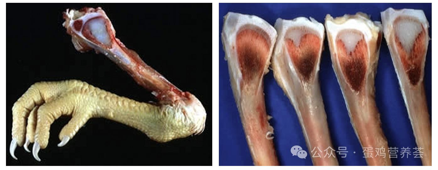
(Left: Tibial dyschondroplasia (chicken) and cartilage plug. In a chick with tibial dyschondroplasia, a cartilage plug is present in the growth plate region. Right: Tibial dyschondroplasia, grading of lesion severity. Lesion severity in tibial dyschondroplasia, with a normal growth plate on the left for comparison. Image source: Jean Sander.)
Symptoms may appear early, but usually first become evident at 14-25 days of age. Affected birds are reluctant to move and, when forced to walk, show a waddling or stiff gait. The disease is caused by an interruption of the normal blood supply to the metaphysis of the proximal growth plate of the tarsometatarsus, preventing nutrient supply and thereby disrupting normal ossification. The abnormal cartilage consists of severely degenerated cells, with atrophied cytoplasm and nuclei; the protein and DNA content of the lesional cartilage is significantly reduced.
The exact cause of tibial dyschondroplasia is unknown. Its incidence can be altered through genetic selection and may be influenced by a major sex-linked recessive gene. An imbalance of dietary electrolytes, especially a chloride level that is too high relative to the levels of other dietary cations, may be a major trigger for many clinical signs. More tibial dyschondroplasia also occurs when dietary calcium is low relative to available phosphorus, or when phosphorus is high relative to calcium.
Treatment measures include adjusting the dietary calcium-to-phosphorus ratio and bringing the dietary electrolyte balance to approximately 240 mEq/kg. However, dietary adjustments rarely result in complete recovery of affected birds. Tibial dyschondroplasia can be prevented by controlling growth rate; however, lighting or feed restriction programs must take into account the economic impact of reduced growth rate. Evidence indicates that replacing some dietary vitamin D₃ with metabolites such as 1,25 (OH)₂D₃ can improve chondrocyte differentiation, thereby reducing the occurrence of this skeletal disease.
3 Caged layer fatigue syndrome
High-producing laying hens housed in cages may occasionally become paralyzed during the peak laying period and afterward. This is caused by spinal cord damage resulting from vertebral fractures. The fractures are caused by impaired calcium flux associated with the high output of calcium into eggshells.
Laying hens can achieve an early laying rate of over 95% for at least 6 months, placing heavy demands on the calcium-flow system between the diet, skeleton, and oviduct. As hens age, medullary bone is renewed daily, and calcium demand drops sharply after ovulation and before calcification of the next eggshell. However, structural bone (cortical and trabecular bone) is not renewed during laying. Over time, the medullary bone, which initially protects the structural bone, gradually becomes dispersed within the medullary cavity. This exposes more of the structural bone surface to the activity of osteoclasts and may eventually lead to osteoporosis. This rarely occurs in floor-raised chickens, suggesting that restricted movement in cages is a potential contributing or associated factor.
Affected hens usually lie on their sides at the back of the cage. In the early stages of paralysis, they appear healthy, and there is often a shelled egg in the oviduct and an active ovary. Death is usually caused by starvation or dehydration because the hens cannot reach feed or water on their own. If moved to the floor, affected hens may recover. To prevent a high incidence of caged layer fatigue, ensure that pullets reach a normal body weight at sexual maturity and provide them with a high-calcium diet (at least 4% calcium) at least 7 days before their first oviposition.
Older caged laying hens are also prone to fractures when removed from cages and transported to processing facilities. During the laying cycle, bone strength cannot be substantially improved without affecting other economic traits (such as eggshell quality). This condition is closely associated with high egg production and a sustained laying cycle (consecutive production of 200–230 eggs).
The diet must provide sufficient calcium and phosphorus to prevent deficiencies. However, feeding a diet with a calcium content of > 2.5% during the young growth stage (<16 weeks) can lead to a high incidence of nephritis, visceral gout, and calcium urate deposits in the ureters, and sometimes to high mortality, especially in cases of infection with infectious bronchitis virus.
By supplying 65%-70% of dietary calcium sources as coarse limestone (diameter 2-4 mm) and the remainder as fine limestone, eggshell strength can be maintained and the trend toward declining bone strength reduced. Coarse calcium sources can meet laying hens’ needs when they are greatest and remain in the gizzard, ensuring calcium supply especially at night, when feeding stops and eggshell calcification demand peaks.
If a fast-acting calcium/calcium phosphate preparation is administered immediately after paralysis is first observed, it will usually be effective. Although it may benefit affected laying hens, it poses a risk to healthy flocks (95%-98%); therefore, treatment decisions depend on the severity of the condition and the proportion of the flock affected.