
Abstract: The mortality and culling rate during the peak egg-laying period of broiler breeders is a key indicator affecting the number of qualified hatching eggs and healthy chicks produced per hen housed. Based on tracking production data from 23 flock batches and post-mortem analyses of dead and culled birds, this article identifies the main lesion types and management-related causes of abnormal mortality and culling rates during the peak period. The analysis found that liver rupture and hemorrhage, prolapse, and feed aspiration are the main factors contributing to increased mortality and culling rates, and are closely associated with management details such as body weight control, light stimulation, and feeding programs. This article proposes that, now that severe infectious diseases are effectively controlled, precise body weight management, scientifically designed lighting programs, an orderly feed increase strategy, and refined on-site operations can significantly reduce mortality and culling rates and improve production efficiency.
Keywords: broiler breeders; peak egg production period; mortality and culling rate; fatty liver hemorrhagic syndrome; refined management
I. Physiological characteristics and challenges during the peak laying period
Broiler breeders enter a period of rapid reproductive system development at 24 to 28 weeks of age, during which metabolism and physiological functions change dramatically. The precision of management during this period directly affects egg production performance and flock health.
Taking Kebo varieties as an example, the changes in the main physiological indicators during the period from 24 to 28 weeks of age are as follows:
Body weight and feed consumption: Cumulative body weight increased by more than 300 grams (an increase of approximately 10%), and weekly feed consumption increased by 50 grams (an increase of approximately 30%), placing greater demands on nutritional supply.
Light stimulation: Light intensity increases from 20 lux to 60 lux, and the duration increases from 8 hours to more than 15 hours. The sharp increase in light intensity strongly stimulates the endocrine system.
Reproductive system development: egg production rate increases by approximately 80%, the distance between the pubic bones widens by 2–3 centimeters, the oviduct length increases from 16 centimeters to 34 centimeters, the ovary weight increases from 71 grams to 148 grams, and egg weight increases by approximately 5 grams.
Hormone level changes: Estrogen and progesterone levels rise rapidly, promoting the maturation of the reproductive system; however, an increase in glucocorticoids may suppress immune function, leading to reduced disease resistance in the flock and an increased risk of infection.
While the changes described above have driven up the egg production rate, they have also significantly increased the physiological burden on the flock and its sensitivity to the environment, bringing the mortality and culling rate into a high-incidence period.
II. Analysis of the Causes of Abnormally High Mortality and Culling Rates During Peak Egg Production
1, patterns of change in mortality and culling rates
Statistical analysis of production data for batches 23 during 2024–2025 years shows:
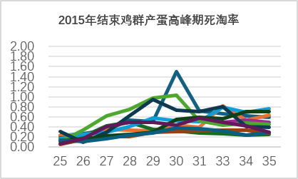
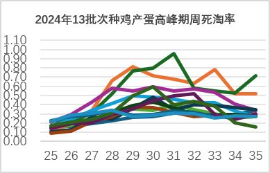
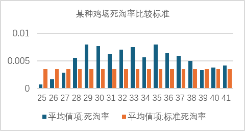
The mortality and culling rate tends to increase as the egg production rate and feed intake increase; this is a general trend.
Based on a weekly mortality and culling rate of 0.3%, the proportions of abnormal batches in 2024 and 2025 were as high as 77% and 80%, respectively.
Mortality and culling rates typically peak at 30 weeks of age. At 35 weeks of age, approximately 70% of batches return to normal levels. However, batches with higher-than-usual mortality and culling rates during the peak period continue to have rates significantly above the standard afterward, as shown in the table below.
2, Classification of lesions found during necropsies of dead and culled chickens
Anatomical analysis of culled chickens aged 25–30 weeks showed the following main types of lesions (a single chicken may exhibit multiple symptoms):
Liver swelling, hemorrhage, and rupture 28%; egg binding (a mature egg in the uterus of a dead hen rules out insufficient blood calcium) 18%; prolapse 64%; peritonitis 25%; choking on feed 45%; intestinal bleeding and diarrhea 43%; and injuries (skin scratches and leg cuts) 10%. Based on the above classification of necropsy lesions, management factors contributing to an increased mortality and culling rate include prolapse, choking on feed, and liver hemorrhage and rupture, as well as secondary bacterial diseases in the later period, such as peritonitis and enteritis.
The relationship between 3, being overweight, liver rupture, and rectal prolapse
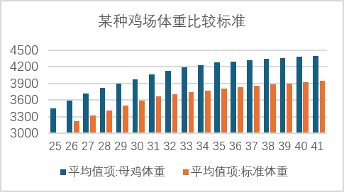
The body weights of chickens that died from liver rupture and prolapse generally exceeded the standard by more than 20%, indicating typical cases of obesity.
Excessive body fat accumulation exceeds the liver’s metabolic capacity, causing damage to the liver tissue structure and making it fragile and prone to breaking. Under stressful conditions such as light stimulation, feed addition, competition for feed, or mating, liver rupture and bleeding can easily be triggered, resulting in death.
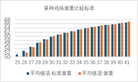
Overweight flocks of chickens are often associated with excessive egg weight and an increased proportion of double-yolk eggs (up to 8%). Double-yolk eggs are difficult to lay, significantly increasing the risk of prolapse, vent pecking, and death from bleeding. The following data show changes in egg weight, mortality, and laying rate caused by excessive body weight.
Deaths and culling caused by improper management practices 4
Feed competition and choking on feed: Feeding immediately after turning on the lights in the early morning, after the flock has fasted for 10 hours, can cause intense excitement, leading to competition for feed, trampling, and running. Feed pellets may accidentally enter the trachea, causing suffocation or neurogenic fainting.
Trauma and secondary infection: Crowding at the feeding area, fighting during mating, and crossing feed lines can cause skin and joint injuries. If not detected and treated promptly, these injuries can develop into systemic infections that are fatal.
(To be continued........)